Your Mild background parenchymal enhancement images are ready. Mild background parenchymal enhancement are a topic that is being searched for and liked by netizens now. You can Get the Mild background parenchymal enhancement files here. Find and Download all royalty-free images.
If you’re searching for mild background parenchymal enhancement pictures information related to the mild background parenchymal enhancement interest, you have pay a visit to the ideal blog. Our site always provides you with hints for seeking the maximum quality video and picture content, please kindly hunt and find more informative video articles and graphics that fit your interests.
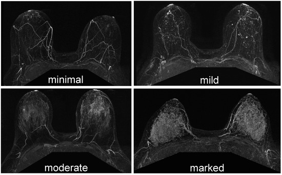
Mild Background Parenchymal Enhancement. Any degree of background parenchymal enhancement beyond baseline was associated with an increased risk of breast cancer development however mammographic breast density and the extent of fibroglandular breast tissue were not associated with any change in baseline breast cancer risk. Background parenchymal enhancement bpe on breast mri refers to the normal enhancement of the fibroglandular tissue and is categorized by the breast imaging reporting and data system birads as minimal mild moderate and marked4moderate and marked bpe can possibly be misinterpreted as suspicious or may mask actual malignant lesions5. Epub ahead of print. BPE may vary in degree and distribution in different patients as well as in the same patient over time.
 Prophylactic Breast Irradiation Reduces Background Parenchymal Enhancement Bpe On Mri A Secondary Analysis Sciencedirect From sciencedirect.com
All accessible MRI examinations from 128 women during a limited time period in 2016 were evaluated. BPE may vary in degree and distribution in different patients as well as in the same patient over time. Mild moderate and marked background parenchymal enhancement is associated with a significantly lower rate of BI-RADS categories 1 and 2 assessments and a significantly higher rate of BI-RADS category 3 assessments than minimal enhancement. Background parenchymal enhancement BPE of the breast via magnetic resonance imaging MRI is associated with future invasive breast cancer risk independent of breast density according to results from a recent study published in the Journal of Clinical Oncology 2019 Jan 9. Epub ahead of print. 101371journalpone0158573 Cite This Page.
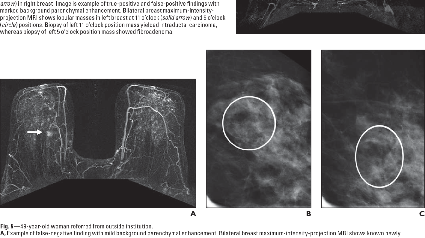
A 55-year-old woman with mild background parenchymal enhancement BPE and affected by invasive ductal carcinoma of the right breast.
They found that the presence of background parenchymal enhancement on a womans initial MRI seemed to predict a greater cancer risk. To investigate if baseline andor changes in contralateral background parenchymal enhancement BPE and fibroglandular tissue FGT measured on magnetic resonance imaging MRI and mammographic breast density MD can be used as imaging biomarkers for overall and recurrence-free survival in patients with invasive lobular carcinomas. A blinded radiologist visually categorized BPE as minimal mild moderate or marked. For each mri examination background parenchymal enhancement was prospectively assigned one of four categories in accordance with the anticipated bi-rads mri lexicon classification system. Moderate or marked background parenchymal enhancement was also associated with a higher abnormal interpretation rate compared with minimal or mild background parenchymal enhancement 305 vs 233. Background parenchymal enhancement BPE is defined as the initial enhancement of the normal breast tissue in the standardized dynamic contrast-enhanced magnetic resonance imaging MRI.
 Source: academicradiology.org
A 55-year-old woman with mild background parenchymal enhancement BPE and affected by invasive ductal carcinoma of the right breast. Endogenous estrogen concentrations are positively. BPE may vary in degree and distribution in different patients as well as in the same patient over time. MRI Background Parenchymal Enhancement Is Not Associated with Breast Cancer. Minimal mild moderate and marked.
 Source: researchgate.net
1a mild 2550 enhancement of glandular tissue fig. BPE does not directly correlate with amount of fibroglandular tissue seen on mammography. The MIP image gives a good overview of both breasts. Background Background parenchymal enhancement BPE on breast magnetic resonance imaging MRI may be associated with breast cancer risk but previous studies of the association are equivocal and limited by incomplete blinding of BPE assessment. Minimal mild moderate and marked.
 Source: jksronline.org
1b moderate 5075 enhancement of. For each mri examination background parenchymal enhancement was prospectively assigned one of four categories in accordance with the anticipated bi-rads mri lexicon classification system. Endogenous estrogen concentrations are positively. 101371journalpone0158573 Cite This Page. Minimal mild moderate and marked.
 Source: sciencedirect.com
1a mild 2550 enhancement of glandular tissue fig. Transverse THRIVE MR image obtained after intravenous administration of a gadolinium chelate in the transverse plane and B subtracted THRIVE MR image show the tumor that was easily detectable as a mass-like lesion. BPE is categorized as minimal mild moderate and marked according to the Breast Imaging Reporting and Data System BI-RADS 1 3. MRI Background Parenchymal Enhancement Is Not Associated with Breast Cancer. Background parenchymal enhancement on breast MRI refers to the normal contrast enhancement of fibroglandular tissue.
 Source: mdpi.com
Women with at least mild background parenchymal enhancement. Background parenchymal enhancement BPE of the breast via magnetic resonance imaging MRI is associated with future invasive breast cancer risk independent of breast density according to results from a recent study published in the Journal of Clinical Oncology 2019 Jan 9. Background parenchymal enhancement is assessed as either symmetric or asymmetric. BPE may vary in degree and distribution in different patients as well as in the same patient over time. Epub ahead of print.
 Source: oncohemakey.com
101371journalpone0158573 Cite This Page. Background Parenchymal Enhancement at Breast MR Imaging. Minimal 25 enhancement of glandular tissue fig. BPE may vary in degree and distribution in different patients as well as in the same patient over time. Normal parenchymal enhancement at breast MR imaging is termed background parenchymal enhancement BPE.
 Source: researchgate.net
Epidemiology Background parenchymal enhancement is more common in younger patients with dense breasts 18. BPE is categorized as minimal mild moderate and marked according to the Breast Imaging Reporting and Data System BI-RADS 1 3. Background parenchymal enhancement BPE of the breast via magnetic resonance imaging MRI is associated with future invasive breast cancer risk independent of breast density according to results from a recent study published in the Journal of Clinical Oncology 2019 Jan 9. Reflecting hormonal influence background enhancement is decreased after menopause 2. Normal Patterns Diagnostic Challenges and Potential for False-Positive and False-Negative.
 Source: semanticscholar.org
MRI-density is a volumetric measure of breast density that is highly correlated with mammographic density an established breast cancer risk factor. Background parenchymal enhancement is assessed as either symmetric or asymmetric. Endogenous estrogen concentrations are positively. Transverse THRIVE MR image obtained after intravenous administration of a gadolinium chelate in the transverse plane and B subtracted THRIVE MR image show the tumor that was easily detectable as a mass-like lesion. Background parenchymal enhancement bpe on breast mri refers to the normal enhancement of the fibroglandular tissue and is categorized by the breast imaging reporting and data system birads as minimal mild moderate and marked4moderate and marked bpe can possibly be misinterpreted as suspicious or may mask actual malignant lesions5.

BPE is categorized as minimal mild moderate and marked according to the Breast Imaging Reporting and Data System BI-RADS 1 3. Normal parenchymal enhancement at breast MR imaging is termed background parenchymal enhancement BPE. They found that the presence of background parenchymal enhancement on a womans initial MRI seemed to predict a greater cancer risk. To investigate if baseline andor changes in contralateral background parenchymal enhancement BPE and fibroglandular tissue FGT measured on magnetic resonance imaging MRI and mammographic breast density MD can be used as imaging biomarkers for overall and recurrence-free survival in patients with invasive lobular carcinomas. This prominence is mostly due to the fact that similar to the association of breast cancer and breast density on.
 Source: file.scirp.org
BPE is thought to be under the effect of blood flow in dense. Normal parenchymal enhancement at breast MR imaging is termed background parenchymal enhancement BPE. Background parenchymal enhancement BPE which represents normal fibro-glandular tissue enhancement in DCE-MRI is considered to relate to hormonally active glandular tissue 2. Transverse THRIVE MR image obtained after intravenous administration of a gadolinium chelate in the transverse plane and B subtracted THRIVE MR image show the tumor that was easily detectable as a mass-like lesion. Normal Patterns Diagnostic Challenges and Potential for False-Positive and False-Negative.
 Source: onlinelibrary.wiley.com
Moderate or marked background parenchymal enhancement was also associated with a higher abnormal interpretation rate compared with minimal or mild background parenchymal enhancement 305 vs 233. BPE may vary in degree and distribution in different patients as well as in the same patient over time. Women with at least mild background parenchymal enhancement. MRI Background Parenchymal Enhancement Is Not Associated with Breast Cancer. Background parenchymal enhancement BPE is defined as the initial enhancement of the normal breast tissue in the standardized dynamic contrast-enhanced magnetic resonance imaging MRI.
 Source: semanticscholar.org
They found that the presence of background parenchymal enhancement on a womans initial MRI seemed to predict a greater cancer risk. There are four terms that describe the BPE. To investigate if baseline andor changes in contralateral background parenchymal enhancement BPE and fibroglandular tissue FGT measured on magnetic resonance imaging MRI and mammographic breast density MD can be used as imaging biomarkers for overall and recurrence-free survival in patients with invasive lobular carcinomas. Reflecting hormonal influence background enhancement is decreased after menopause 2. PLOS ONE 2016.
 Source: semanticscholar.org
Background parenchymal enhancement BPE which represents normal fibro-glandular tissue enhancement in DCE-MRI is considered to relate to hormonally active glandular tissue 2. BPE may vary in degree and distribution in different patients as well as in the same patient over time. This prominence is mostly due to the fact that similar to the association of breast cancer and breast density on. Background parenchymal enhancement BPE is defined as the initial enhancement of the normal breast tissue in the standardized dynamic contrast-enhanced magnetic resonance imaging MRI. Background parenchymal enhancement on breast MRI refers to the normal contrast enhancement of fibroglandular tissue.
 Source: researchgate.net
Mild moderate and marked background parenchymal enhancement is associated with a significantly lower rate of BI-RADS categories 1 and 2 assessments and a significantly higher rate of BI-RADS category 3 assessments than minimal enhancement. We investigated the correlations between background parenchymal enhancement BPE and MRI interpretations with respect to short-interval follow-ups and biopsy rates. This prominence is mostly due to the fact that similar to the association of breast cancer and breast density on. Imaging is termed background parenchymal enhancementBPE. Normal Patterns Diagnostic Challenges and Potential for False-Positive and False-Negative.

Normal parenchymal enhancement at breast MR imaging is termed background parenchymal enhancement BPE. BPE may vary in degree and distribution in different patients as well as in the same patient over time. Women with at least mild background parenchymal enhancement. MRI Background Parenchymal Enhancement Is Not Associated with Breast Cancer. Moderate or marked background parenchymal enhancement was also associated with a higher abnormal interpretation rate compared with minimal or mild background parenchymal enhancement 305 vs 233.
 Source: healthmanagement.org
They found that the presence of background parenchymal enhancement on a womans initial MRI seemed to predict a greater cancer risk. We investigated the correlations between background parenchymal enhancement BPE and MRI interpretations with respect to short-interval follow-ups and biopsy rates. Endogenous estrogen concentrations are positively. Background parenchymal enhancement BPE which is defined as the enhancement of normal fibroglandular tissue on contrast-enhanced dynamic breast magnetic resonance imaging MRI has drawn considerable attention in recent years. BPE is categorized as minimal mild moderate and marked according to the Breast Imaging Reporting and Data System BI-RADS 1 3.
 Source: onlinelibrary.wiley.com
They found that the presence of background parenchymal enhancement on a womans initial MRI seemed to predict a greater cancer risk. Women with at least mild background parenchymal enhancement. Background parenchymal enhancement BPE which represents normal fibro-glandular tissue enhancement in DCE-MRI is considered to relate to hormonally active glandular tissue 2. Background parenchymal enhancement is assessed as either symmetric or asymmetric. Background Parenchymal Enhancement at Breast MR Imaging.
 Source: semanticscholar.org
Background parenchymal enhancement is assessed as either symmetric or asymmetric. A blinded radiologist visually categorized BPE as minimal mild moderate or marked. Mild moderate and marked background parenchymal enhancement is associated with a significantly lower rate of BI-RADS categories 1 and 2 assessments and a significantly higher rate of BI-RADS category 3 assessments than minimal enhancement. Women with at least mild background parenchymal enhancement. Moderate or marked background parenchymal enhancement was also associated with a higher abnormal interpretation rate compared with minimal or mild background parenchymal enhancement 305 vs 233.
This site is an open community for users to do sharing their favorite wallpapers on the internet, all images or pictures in this website are for personal wallpaper use only, it is stricly prohibited to use this wallpaper for commercial purposes, if you are the author and find this image is shared without your permission, please kindly raise a DMCA report to Us.
If you find this site helpful, please support us by sharing this posts to your preference social media accounts like Facebook, Instagram and so on or you can also save this blog page with the title mild background parenchymal enhancement by using Ctrl + D for devices a laptop with a Windows operating system or Command + D for laptops with an Apple operating system. If you use a smartphone, you can also use the drawer menu of the browser you are using. Whether it’s a Windows, Mac, iOS or Android operating system, you will still be able to bookmark this website.


